Hysteroscopic surgery for intrauterine diseases
Specialist:
Peng-Hui Wang
Yi-Jen Chen
Yen-Hou Chang
Chia-Hao Liu
I-San Chan
Wei-Ting Chao
Overview
Operative hysteroscopy is indicated for the treatment of various conditions such as abnormal uterine bleeding, submucosal leiomyoma, uterine polyps, uterine septum, uterine adhesion, or blockage of the fallopian tubes.
Intrauterine diseases are characterized by pathological lesions, such as endometrial polyps, submucosal myomas, or uterine synechiae, or congenital malformations of the uterus, such as a bicornuate or septate uterus. Diagnostic hysteroscopy is the most direct way to visualize and evaluate the interior of the uterus. When patients with intrauterine diseases experience abnormal vaginal bleeding, spontaneous abortion, or infertility, an operative hysteroscopy may be a therapeutic treatment option.
Features
Operative hysteroscopy can serve to insert ancillary devices such as scissors or graspers to perform surgical procedures inside the uterine cavity and assist with specific procedures like cannulation of the fallopian tube under hysteroscopy. A specialized electrosurgical hysteroscope, called a resectoscope, is commonly used to perform the resection of endometrial polyps, uterine myomas, or uterine septa.
An innovative hysteroscopic tissue removal system has been developed based on the mechanical removal of intrauterine lesions. This system doesn't require a bipolar system and causes less injury to the uterus. The tissue is collected through the fluid delivery system, which filters the specimen into a bag. The cost is expected to increase with the new device.
Procedure
- The patient is placed in a lithotomy position and given anesthesia.
- The cervical canal is carefully dilated with Hegar dilators, and the operative hysteroscope is inserted into the uterine cavity under direct vision.
- During the procedure, a media, usually normal saline, is instilled to distend the uterine cavity adequately and allow the cavity to be scanned with all landmarks.
- The operative procedure is performed through the system of the operative hysteroscope.
- In some cases, simultaneous laparoscopy may be required during specific situations.
Notification
The operative hysteroscopy procedure carries risks, including:
- Distending media-related adverse events: excessive fluid absorption (fluid overload) and electrolyte imbalance.
- Uterine perforation.
- Intra-operative bleeding.
- Thermal injury, which may occur at adjacent sites such as the cervix, vagina, and vulva and may also occur to intraperitoneal structures like the bowel if a uterine perforation occurs.
- Endometrial or myometrial infection.
- Post-operative intrauterine adhesion.
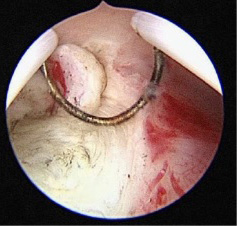
A cutting loop is used to remove the myoma stalk
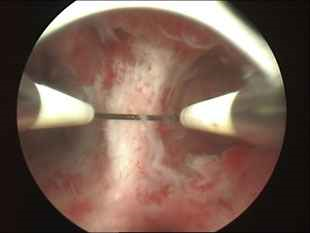
The cutting loop is used to resect the uterine septum.
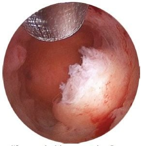
The mechanical morcellator is used to remove the endometrial polyp.
Last Modified: